A major challenge in the field of biosesning is the detection of trace amounts of target proteins in biological samples. Fiber-optic biosensors present an affordable technique for target protein bio-detection with great sensitivity. However, their specificity is limited. This proposal suggests developing a novel microfiber biosensor, which is significantly enhanced by the ability to detect the amount and location of single gold nanoparticles via photothermal phase imaging as shown in Fig.1.
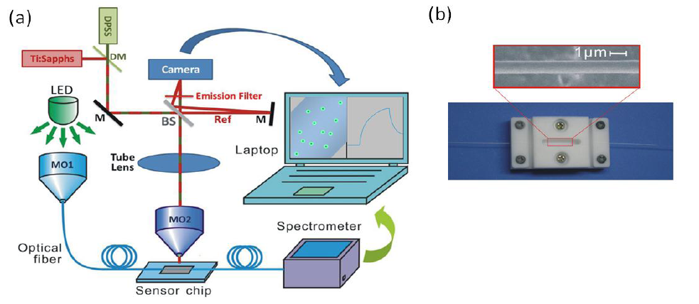
Fig.1 (a) Illustration of the proposed experimental setup, (b) Image of a sensor cell and scanning electron microscopy image of the optical microfiber
Based on the previous knowledge and experience of both groups, Prof.Wu’s group specialized in microfiber biosensors, including their special fabrication and biological uses. During the project, a gold nanopariticle amplified optical microfiber evanescent wave absorption biosensor for cancer biomarker detection was proposed. As depicted in Fig.2(a), a sandwich assay was used for the detection of the protein molecules. Besides, a single nano-magnetic bead or nanorod can be detected using nanofiber biosensor shown in Fig.2(b) &(c). To further improve the sensitivity of this biosensor, the tapered fiber is coupled to a whispering-gallery-mode resonator, which is expected to achieve single biomolecule detection.
(a)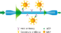 (b)
(b)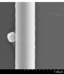 (c)
(c)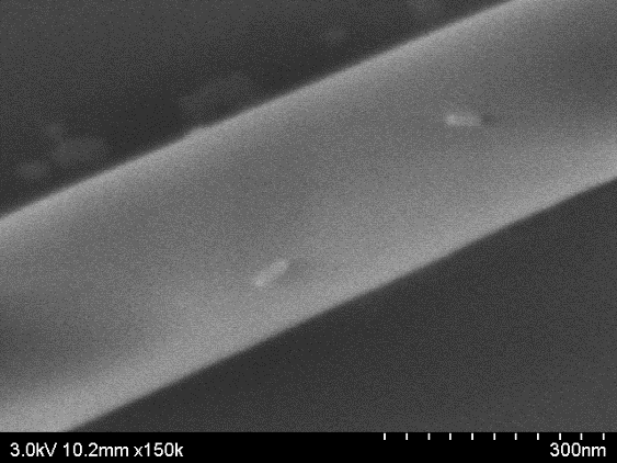
Fig.2 (a)Schematic diagram of the immunoassay using the GNPs as signal amplification labels, (2) A single nano-magnetic bead on the fiber surface, (3) A single nanorod on the fiber surface
On the other hand, Prof.Shaked’s group specialized in interferometric phase microscopy and nanoscopy. To predict the spatial phase signal from photothermally excited metallic nanoparticles of the arbitrary shapes and sizes, a new approach based on a finite-volume method was developed. And various shapes of nanoparticles are simulated using this model and verified with experimental results. Meanwhile, two-wavelength phase unwrapping using external module for multiplexing off-axis holography was proposed to achieve quantitatively imaging.
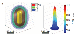
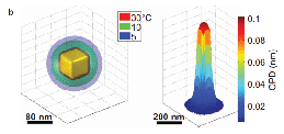
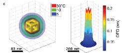
Fig.3 Demonstration of the use of the proposed model to simulate the thermal and OPD distributions of gold nanoparticles of various shapes.
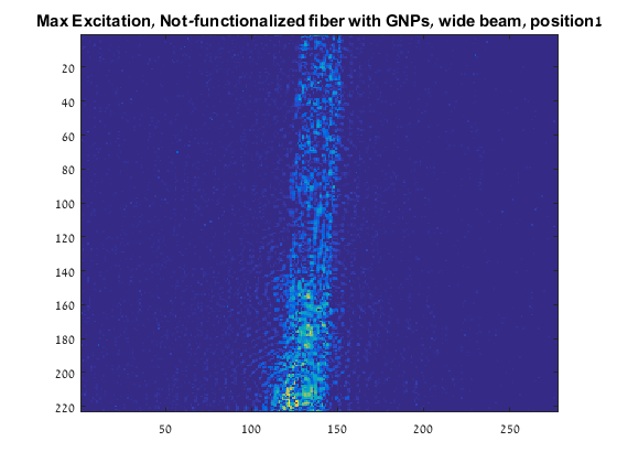
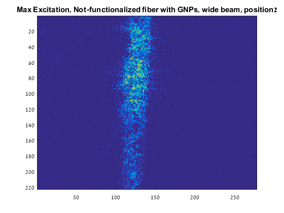
Nowadays, nanoparticles can be detected using this photo-thermal interferometric phase microscopy, as displayed in Fig.4. It is obvious that the 50nm nanoparticles on the fiber can be recognized.
In future, we will continue to target protein and nucleic acid bio-detection combing the photothermal interferometric phase microscopy with the nanofiber biosensor for cancer detection.

 中文
中文